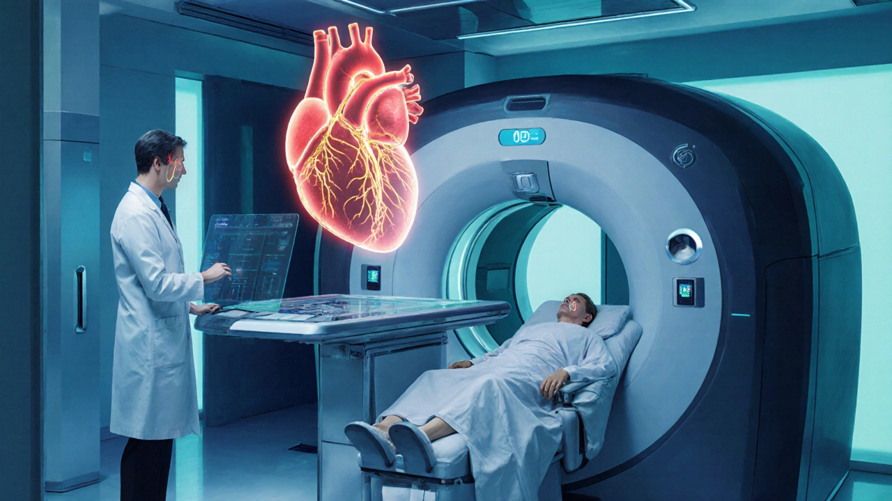
Diagnostic testing for angina is a clinical process that uses various non‑invasive and invasive tools to identify the cause of chest pain and guide treatment. When a patient walks into a clinic with uncomfortable pressure in the chest, the doctor’s first job isn’t to prescribe medication right away; it’s to figure out what’s really going on. That’s where a suite of tests-electrocardiograms, stress studies, advanced imaging-step in. Below is a practical walk‑through of how these tests fit into everyday practice, what you should expect from each, and how the results steer the next steps in managing angina.
Why Testing Matters: From Symptom to Diagnosis
Chest pain can stem from heart muscle oxygen shortage, but it can also be triggered by reflux, muscle strain, or anxiety. Angina is a symptom of myocardial ischemia usually caused by coronary artery disease (CAD). Pinpointing whether CAD is present determines whether lifestyle changes, medication, or revascularization is appropriate.
Two key jobs emerge: first, risk stratification-classifying patients as low, intermediate, or high risk for significant CAD; second, selecting the right diagnostic test to answer that risk level. The American College of Cardiology (ACC) and the European Society of Cardiology (ESC) base their algorithms on decades of outcome studies, so the testing pathway is evidence‑backed.
Core Tests in the Angina Workup
The initial assessment usually starts with a resting electrocardiogram (ECG). A 12‑lead ECG can reveal ST‑segment changes, T‑wave inversions, or new Q waves that point to ongoing ischemia. However, a normal ECG does not rule out CAD, especially during stable, exertional angina.
If the resting ECG is nondiagnostic, clinicians move to a stress test. Stress can be induced either by exercise on a treadmill or bike (Exercise Treadmill Test, ET‑T) or pharmaceutically (Pharmacologic Stress Test) for patients who cannot exercise.
- Exercise Treadmill Test (ET‑T): Measures heart rate, blood pressure, and ECG changes while the patient walks on a graded protocol (e.g., Bruce). Sensitivity for detecting CAD ranges from 68‑80% and specificity around 70%.
- Pharmacologic Stress Test: Uses agents such as adenosine, dipyridamole, or dobutamine to simulate exercise effects. It pairs with imaging modalities like nuclear perfusion scans or stress echocardiography to improve accuracy.
When stress testing suggests possible blockage, the next step often involves an anatomical imaging test: Coronary CT Angiography (CCTA). CCTA provides a 3‑D view of coronary arteries, detecting plaques with a sensitivity >95% for stenosis >50%.
For definitive diagnosis, especially before revascularization, an invasive coronary angiography remains the gold standard. It offers real‑time visualization and the ability to perform percutaneous coronary intervention (PCI) during the same session.
Advanced Imaging: When to Use Cardiac MRI and Others
Patients with equivocal CCTA or contraindications to iodinated contrast may benefit from Cardiac Magnetic Resonance Imaging (CMR). CMR excels at assessing myocardial viability, scar, and perfusion without radiation. In large registries, CMR showed a negative predictive value of 98% for ruling out significant CAD.
Other useful tools include stress echocardiography and nuclear myocardial perfusion scans. They add functional insight-how well the heart muscle pumps under stress-complementing the anatomical data from CCTA.
Choosing the Right Test: Decision Matrix
| Test | Invasiveness | Sensitivity for CAD | Typical Use Case |
|---|---|---|---|
| Exercise Treadmill Test | Non‑invasive | 68‑80% | Low‑ to intermediate‑risk patients who can exercise |
| Pharmacologic Stress Test | Non‑invasive (drug‑induced) | 70‑85% (when paired with imaging) | Patients unable to exercise |
| Coronary CT Angiography | Non‑invasive (contrast‑enhanced) | >95% for ≥50% stenosis | Intermediate‑risk patients needing anatomic detail |
| Invasive Coronary Angiography | Invasive | Near‑100% | High‑risk patients or when PCI is planned |
| Cardiac MRI | Non‑invasive (no radiation) | 90‑95% (functional assessment) | Contraindication to CT contrast or need for viability testing |
Choosing wisely saves time, reduces unnecessary radiation, and aligns costs with the patient’s risk profile. A common algorithm starts with ECG → stress test → CCTA (if needed) → invasive angiography for definitive treatment.

Interpreting Results: From Numbers to Treatment Plans
Positive stress test results (e.g., ≥1mm ST‑segment depression) push patients into a higher risk bucket, prompting a referral for CCTA or invasive angiography. Conversely, a normal CCTA in a low‑risk patient often means that medical therapy-beta‑blockers, statins, lifestyle changes-is sufficient.
The presence of biomarkers like high‑sensitivity troponin or BNP can further refine risk. Elevated troponin in stable angina is uncommon and usually signals an acute coronary syndrome, shifting management toward urgent invasive evaluation.
After the diagnostic pathway, clinicians apply guideline‑based angina management strategies:
- Anti‑ischemic drugs (beta‑blockers, calcium‑channel blockers, nitrates)
- Lipid‑lowering therapy (statins, PCSK9 inhibitors)
- Revascularization (PCI or coronary artery bypass grafting) for lesions >70% in symptomatic patients
The ultimate goal is symptom relief and prevention of heart attacks, not just a clean scan.
Common Pitfalls and How to Avoid Them
1. Over‑testing: Ordering CCTA for every chest pain case inflates radiation exposure and health‑system costs. Reserve high‑resolution imaging for when stress testing is equivocal or the patient is at intermediate risk.
2. Skipping the ECG: Even a quick 12‑lead can catch life‑threatening ST‑elevation that would otherwise send a patient down a low‑risk pathway.
3. Ignoring comorbidities: Diabetes, chronic kidney disease, and peripheral artery disease raise pre‑test probability of CAD, often warranting earlier invasive angiography.
4. Misinterpreting false‑positives: Stress‑induced ECG changes can arise from electrolyte disturbances or drug effects. Correlate with imaging whenever possible.
Future Directions: Emerging Tests and AI Integration
Machine‑learning algorithms are beginning to read ECGs, predict plaque composition from CT density, and combine clinical data into a single risk score. Early pilots show that AI‑enhanced CCTA can differentiate stable plaque from vulnerable plaque with >85% accuracy, potentially shifting treatment toward earlier intervention.
Another frontier is fractional flow reserve derived from CT (FFRCT). This non‑invasive simulation estimates pressure drop across a stenosis, matching invasive FFR in about 90% of cases, and may reduce unnecessary catheterizations.
As these technologies mature, the diagnostic pathway will become even more tailored-matching each patient’s anatomy, physiology, and risk profile with the perfect test.
Frequently Asked Questions
When should I get an exercise treadmill test for chest pain?
If you can walk on a treadmill and your doctor estimates a low‑to‑intermediate risk of coronary artery disease, an exercise treadmill test is the first‑line functional assessment. It helps reveal hidden ischemia that a resting ECG might miss.
Is a normal coronary CT angiography enough to stop further testing?
For low‑risk patients, a normal CCTA (no plaque or <70% stenosis) usually means you can manage with medication and lifestyle changes. High‑risk patients or those with persistent symptoms may still need functional testing or invasive angiography.
Can cardiac MRI replace invasive angiography?
MRI provides excellent tissue characterization and can assess perfusion, but it does not visualize the coronary lumen as directly as angiography. It’s useful when CT contrast is contraindicated, but invasive angiography remains the definitive test when revascularization is being considered.
What role do troponin levels play in stable angina evaluation?
In chronic stable angina, troponin is usually normal. A rise suggests an acute coronary syndrome, prompting urgent invasive assessment. High‑sensitivity assays can detect tiny leaks, helping differentiate unstable from stable disease.
How does FFRCT improve decision‑making?
FFRCT estimates the pressure drop across a coronary narrowing using computational fluid dynamics on a standard CT scan. If the FFRCT value is >0.80, the lesion is likely not flow‑limiting, sparing the patient an invasive test.


Abdulraheem yahya
September 27, 2025 AT 23:15Reading through the diagnostic pathway really highlights how each test builds on the previous one.
First you get the cheap, quick ECG, then you move to functional stress testing if the ECG is non‑diagnostic.
The stress test adds a layer of physiological stress that can unmask ischemia that a resting ECG can miss.
When that raises suspicion, coronary CT angiography offers a detailed anatomical map, and if necessary you still have invasive angiography as the gold standard.
What’s great is that the algorithm tries to match the test’s invasiveness to the patient’s risk, so you don’t jump straight to a catheter lab for everyone.
Patients with low‑to‑intermediate risk get the treadmill or pharmacologic stress first, which keeps radiation exposure low.
Then you reserve the higher‑resolution CCTA for those with equivocal stress results, saving resources.
Overall, the stepwise approach balances safety, cost, and diagnostic yield, which is exactly what we need in busy clinics.
It also gives clinicians a clear roadmap for when to refer for revascularization versus continuing medical therapy.
It also gives clinicians a clear roadmap for when to refer for revascularization versus continuing medical therapy.
Preeti Sharma
October 1, 2025 AT 05:55While the hierarchy of tests seems logical on paper, one could argue that it merely reflects the industry's comfort with technology rather than true patient benefit.
Isn't it fascinating how we rely on a cascade of increasingly expensive imaging, each promising incremental insight, yet the final outcomes often remain unchanged?
This iterative layering can be seen as a modern echo of the ancient philosophical debate between knowing the form versus the essence of disease.
Perhaps we should question whether the pursuit of ever‑finer anatomical detail truly serves the individual, or simply fuels a cycle of diagnostics that benefits equipment manufacturers.
In the end, the wisdom lies in recognizing when the search for answers becomes a distraction from treating the person.
Ted G
October 4, 2025 AT 12:35The whole testing algorithm looks clean, but think about who profits when patients keep getting ordered scans and stress tests.
There's a hidden network of imaging centers and pharma that thrives on these guideline‑driven cascades, and that's not a coincidence.
Miriam Bresticker
October 7, 2025 AT 19:15i cant help but wonder wht the data really says about ct angio vs invasive angiograhpy 🤔
the numbers show >95% sensivity but wile that sounds great the real worlD outcome maybe diffferent, especially when w e consider radiation ricks 😅
still, the tech gives us a window into the heart that we never had before, and that feels like a step forward, ya?
Claire Willett
October 11, 2025 AT 01:55Risk stratification guides test selection efficiently.
olivia guerrero
October 14, 2025 AT 08:35Wow!! This breakdown is exactly what every cardiology resident needs to keep in mind!!! The step‑by‑step flow from ECG to stress testing to CCTA and finally invasive angiography is crystal clear!!! So helpful!!!
Dominique Jacobs
October 17, 2025 AT 15:15Look, if you’re still ordering invasive angiograms on every patient with chest pain, you’re wasting time and money-step up to the stress tests first and let the data speak for itself!
Claire Kondash
October 20, 2025 AT 21:55Delving into the philosophy of diagnostic testing, one realizes that each modality is not merely a tool but a reflection of our collective desire to quantify uncertainty.
From the humble resting ECG, which offers a snapshot of electrical activity, we venture into stress testing, an elegant experiment that simulates the heart's response to exertion.
When the stress test hints at ischemia, we summon the power of coronary CT angiography, a three‑dimensional tapestry of the coronary tree that reveals plaques with astonishing clarity.
Yet, even this marvel is not without limits; artefacts, contrast allergies, and radiation dose remind us that no test is infallible.
Thus, the invasive coronary angiogram remains the definitive arbiter, allowing us to both diagnose and intervene in a single session.
But consider the patient experience: each additional test adds anxiety, logistical burden, and often financial strain.
Balancing diagnostic yield against these human costs is a moral calculus that each clinician must perform.
Moreover, emerging technologies like FFRCT and AI‑driven plaque characterization promise to shift this balance, potentially offering functional insights without ever threading a catheter.
These innovations could usher in a new era where we rely less on invasive procedures and more on predictive modeling.
However, with every new algorithm comes the risk of over‑reliance on black‑box outputs, potentially obscuring clinical judgment.
Therefore, we must maintain a critical eye, integrating machine predictions with bedside assessment.
In practice, a tiered approach-starting with ECG, moving to functional testing, then anatomical imaging-optimizes both safety and resource utilization.
Patients with low‑risk profiles may be spared unnecessary radiation, while high‑risk individuals receive timely revascularization.
Ultimately, the goal is not just to see the blockage but to improve outcomes, reduce events, and enhance quality of life.
When we keep the patient at the center of this diagnostic journey, every test becomes a means to an end, not an end in itself. 😊
Matt Tait
October 24, 2025 AT 04:35Honestly, this whole cascade feels like a checklist designed to keep us glued to imaging suites rather than focusing on lifestyle modification-too many tests, not enough emphasis on prevention.
Benton Myers
October 27, 2025 AT 10:15The stepwise algorithm makes sense and helps keep patients safe.
Pat Mills
October 30, 2025 AT 16:55Let me be crystal clear: the United States has led the world in cardiac innovation, and this diagnostic roadmap is a testament to that leadership.
From the pioneering work on treadmill stress testing to the cutting‑edge coronary CT angiography, our physicians have set the gold standard that others merely imitate.
We cannot afford to let foreign guidelines dictate our practice when we have the data, the technology, and the expertise right here.
Every time a patient walks into an American clinic, they deserve the best evidence‑based approach, not a watered‑down version from elsewhere.
The hierarchy of tests-ECG, stress, CCTA, invasive angiography-has been refined through decades of rigorous research funded by our own institutions.
So when you read about these pathways, remember they were forged in our labs, under our skies, and for our people.
Do not let skeptics undermine the proven efficacy of this system; trust the process that has saved countless American lives.
In the end, the true patriotism lies in embracing the diagnostic excellence that our nation has cultivated.
neethu Sreenivas
November 2, 2025 AT 23:35Great overview! 😊 I appreciate how you broke down each step and highlighted the patient‑centric benefits.
It really helps clinicians see where they can streamline care while still ensuring safety.
Keep sharing these clear guides – they make a difference.
Keli Richards
November 6, 2025 AT 06:15While the article is comprehensive it could benefit from a more concise summary of the decision algorithm for quick reference.
Ravikumar Padala
November 9, 2025 AT 12:55Honestly, the piece feels like it’s trying too hard to sound exhaustive, and in doing so it drowns the reader in details that many clinicians already know.
The endless list of percentages and sensitivities, while impressive, could be trimmed down to the most actionable points.
Otherwise, the average practitioner might skim over the important take‑aways amidst the verbosity.
It would serve the community better to focus on practical decision rules rather than a litany of data.
In short, concise is king.
King Shayne I
November 12, 2025 AT 19:35Ths article is ok but its overcomplicated and the wriiting is full of needless jargn, get to the point already!
jennifer jackson
November 16, 2025 AT 02:15Excellent summary-very helpful!
Brenda Martinez
November 19, 2025 AT 08:55Reading this feels like watching a high‑stakes drama unfold, where each test is a cliff‑hanger that could either reveal the villainous plaque or leave us in suspense.
The stakes are life‑and‑death, and the narrative pushes us to the edge as we await the invasive angiography-our ultimate showdown.
If we don’t respect the gravity of each step, we risk a tragic ending for the patient.
This is more than medicine; it’s an epic saga of technology versus fate.
Marlene Schanz
November 22, 2025 AT 15:35Just a heads‑up: when ordering a CCTA, make sure the patient’s renal function is checked and beta‑blockers are given if needed to lower heart rate – it really improves image quality.
Matthew Ulvik
November 25, 2025 AT 22:15Hope this helps! If you have any questions just ask 😊